![]() Go to frame view (Recommended only for
screen resolution 1024x768)
Go to frame view (Recommended only for
screen resolution 1024x768)
6.10 Automation of the Nucleic Acid Sequencing Process
In 1986-87, the first papers were published describing successful attempts to automate DNA sequencing. The major factor of success was the automation of the gel autoradiogram reading procedure. In this case, fluorescent labels are used instead of radioactive ones, which drastically cuts down the time necessary to detect the oligo(poly)nucleotides separated in the sequencing gel. Moreover, macromolecules are detected in the gel during electrophoresis, and all the acquired data are immediately sent to the computer for storage. In other words, all manual operations to dry the gel, place the X-ray film, expose it and analyze the autoradiograms are eliminated.
The chemistry underlying the method under consideration will be first
illustrated using a very simple technique recommended for enzymatic sequencing as an
example. The primer in the M13 phage system is universal, with an
amino-fluorescein-containing fragment being incorporated into the terminal 5' phosphate in
several steps ![]() .
.
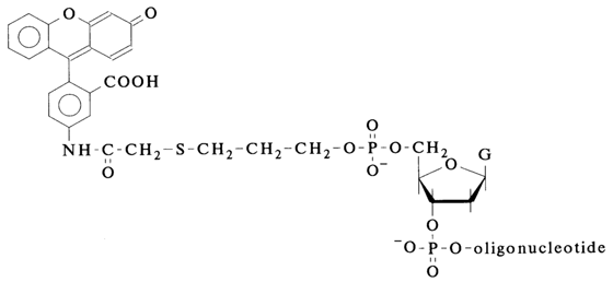
The resulting derivative of the primer oligonucleotide, containing the fluorescent label, is purified by means of ion-exchange high performance liquid chromatography (HPLC), desalted by dialysis and stored in the dark at -200C.
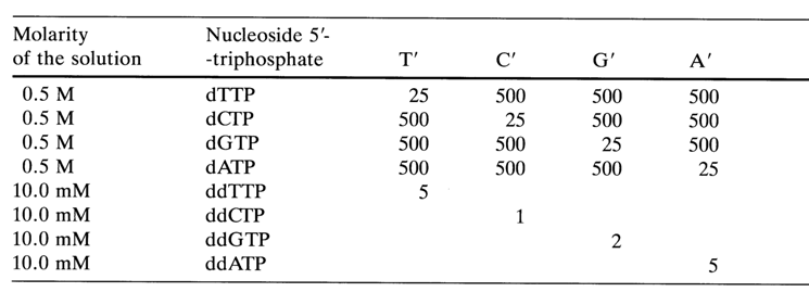
The sequencing is performed as usual but with a slightly altered dNTP:ddNTP ratio. Table 6-7 lists the corresponding quantities (in ml) of the dNTP and ddNTP solutions introduced into the reaction in the case of preparation of terminating mixtures (T', C', G' and A') for determining the positions of T, C, G and A, respectively, in the DNA under analysis.
The presence of a rather sizable fluorescent group in the primer does not affect the process of hybridization with the single-stranded M13 phage DNA.
As has already been mentioned, electrophoresis is performed using a standard procedure with the difference that connected to the instrument is an air-cooled argon laser, light collecting and imaging optics, a photomultiplier (detector), and a data acquisition system interfaced with a computer. Figure 6-34 shows schematically a system for automatic reading of DNA sequencing data obtained during the electrophoresis (i.e. from sequencing gel). The plates for electrophoresis are made of non-fluorescent glass. The sensitivity for detecting macromolecules with a fluorescent label is 3. 10-18 moles/ band. Within mere five hours one can read a sequence of 400 nucleotides (up to a maximum of 500 nucleotides) within a 20 to 30 cm long gel. The system may be used to handle anywhere from six to ten DNA samples per run (gel). The gel may be prepared from both polyacrylamide and agarose.
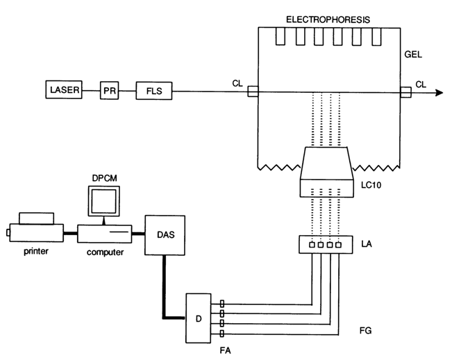
Fig. 6-34. Schematic diagram of the instrument for reading out electrophoresis data during DNA sequencing by the enzymatic method with a fluorescent primer (aminofluorescein). PR - polarization rotator; FLS - focusing lens system; CL - plates for coupling light into gel; LCIO - light collecting and imaging optics; LA - limiting aperture; LG - light guides; FA - filter assembly; D - detector (photomultipliers); DAS - data acquisition system; DPCM - data processing and control module (IBM PC).
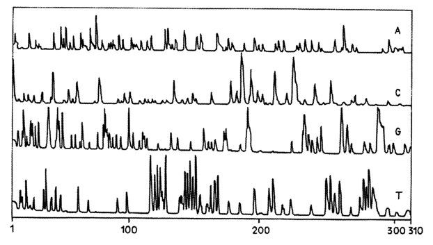
Fig. 6-35. Results of analysis of a sequencing gel (20 cm, 7 % polyaerylamide gel), obtained in enzymatic sequencing of recombinant M13 mp8 (3SV DNA with an aminofluorescein containing primer. The sequence was read from the gel using laser-induced fluorescence (for description of the instrument, see above).
Figures 6-35 and 6-36 illustrate a partial nucleotide sequence of recombinant M13mp8
bSV DNA containing a complex gene composed of rabbit b-globin genes and SV40 inserted into the restriction nuclease AccI and EcoRI sites of vector M13mp8.Among the major advantages of this method, which was developed at the European Molecular Biology Laboratory (EMBL), are its simplicity, high speed and low cost. In spite of the advances in sequencing, associated with radioactive labels, serious difficulties are encountered when using the latter, including hazards in handling, storage and disposal of the waste (labeled products), high cost of radiolabeled compounds, as well as the extremely short lifetime of the 32P-labeled compound usually employed in sequencing.

Fig. 6-36. Excerpt from Fig. 6-35. Shown here is a nucleotide sequence in a rabbit
b-globin gene between positions 189 and 153, which corresponds to bases 140-176 read from the gel.Replacement of radioactive labels by fluorescent labels renders analysis of macromolecules simpler and less costly. The described instrument for continuous reading of sequencing gel data is rather simple and makes it possible to handle many DNA samples within a short period of time.
The company "Applied Biosystems" has started production of automatic DNA sequencing instruments. The procedure for analysing oligo(poly)nucleotides is similar to the one just described, except that the detection of products of each of the four reactions during sequencing by the Sanger's method requires use of four structurally different fluorescent dyes (with four different fluorescence maxima) covalently bound to the primer oligonucleotide. Thus, distinction can be made between the sets of four reactions, which can be combined within a single cell, rather than being conducted in four as in the basic procedure described above. When four different fluorescent labels are selected, the following criteria must be met: (1) the fluorescence spectrum maxima must be clearly distinguishable: (2) all dyes must ensure almost the same high detection sensitivity (ca. 10-18 M); (3) their presence in the primer oligonucleotide should not affect the hybridization of the latter with DNA; and (4) the electrophoretic mobilities of the fluorescently marked DNA fragments should not differ widely because of the dissimilar structures of the fluorescent compounds. The following compounds were used by the authors as fluorescent labels more or less satisfying the above criteria:
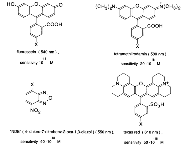
The instrument for direct reading from the gel (electrophoresis is conducted in 50 cm long tubes with 8 % polyacrylamide gel is similar to the one described above. The laser beam passing through the gel at a distance of 40 cm from the top excites fluorescence which is then detected with the aid of four filters selected to match the spectra of each of the four dyes and changing periodically at a fast speed. More specifically, the fluorescence of the abovelisted four dyes was detected at four wavelengths: 520, 550, 580 and 610 nm.
However, in spite of the successful implementation of such an approach for analysis of rather extended DNA fragments, which makes it possible to read simultaneously 16 gel tubes over a period of eight to ten hours, this sequencing procedure based on the automatic instrument of "Applied Biosystems" has some drawbacks. The fluorescence spectra of the dyes partially overlap so that the peaks corresponding to each dye appear in other channels as well. Moreover, the electrophoretic mobility of the labeled fragments varies depending on the label structure. For example, samples labeled with fluorescein and tetramethylrhodamine move in the gel so as if their length exceeds that of the NBD-labeled samples by one monomer, whereas samples labeled with Texas red appear to be one and a quarter monomers longer. In order to obviate such difficulties, special programs of a high degree of sophistication are employed using empirically selected correction factors. For all the attractiveness of the "a-label-for-each-type-of-nucleotide" approach, the method described at the beginning of this section - that is, the one involving a single dye species - is more likely to be universally used. It may be expected that in the near future scientists and companies working on such sequencers will concentrate their efforts on improvement of the existing designs, simplification of the chemical procedures for preparing samples for DNA and RNA analysis, complete automation of the entire sequencing process, and development of sequencers based on the Maxam-Gilbert method as well. The latter offers a number of advantages over Sanger's method, stemming, to be particular, from the use of less expensive reagents, more uniform intensity of the bands corresponding to all reaction products, and the fact that the analysis results are free of any influence exerted by the secondary structure of the DNA being sequenced. It is also possible that new, interesting results from the standpoint of full automation may be produced by combining the solid-phase technique with sequencing based on non-radiolabeled samples.
Doubtless, the automatic sequencing methods already in existence can already be used for determining the primary structure of DNA.
An alternative approach based on a fast, reliable and inexpensive DNA sequencing procedure has been proposed by Japanese scientists and companies belonging to the National Committee for Automation of DNA Sequencing. The general idea brought forth by the Committee boils down to setting up a facility performing assembly-line analyses of the primary DNA structure with the aid of automatic sequencers and robots. Individual components of the automated line for such analysis have already been developed. The process is based on Sanger's polymerase copying method which allows nucleotide sequences to be read at a rate of 0.28 second per base, in contrast to the Maxam-Gilbert method with its rate of 144 seconds per base (automatic sequencers operating on this principle are produced by "Seiko"). Technology has been elaborated for automatic production of ready-to-use polyacrylamide gel slabs (0.02 x 20 x 40 cm) wrapped in an acetate film which permits autoradiography to be performed immediately after electrophoresis. This enables one to get rid of the rather tedious and time-consuming manual operations of preparing gels and drying them after electrophoresis.
Electrophoresis can be sped up by designing units for simultaneous handling of hundreds of gels (each making it possible to analyze five DNA fragments about 250 bases long).
Autoradiograms are read using automatic scanning devices which ensure a rate of 1.4 seconds per base. To reduce the probability of a reading error (which is typically 1 % per 250 bases), one can read two complementary strands at a time, thereby the error probability is brought down to 10-4 . There is every reason to expect faster and less costly analyses if the radioactive label is replaced by a fluorescent one.
The advantages offered by a well organized line for nucleic acid sequence analyses include high speed of the entire process, lower labor requirements, the possibility to maintain high quality of the analyses, ensure their reproducibility and, as a consequence, high reliability of the results. According to the Committee's estimates, at present a single skilled research worker can manually read a thousand base pair long sequence per day at a cost of a dollar per base. Through automation one can attain a rate of a million bases per day (per automatic unit). Such an increase in analysis rate must lower the cost by one order of magnitude.